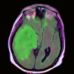
A genuine U-Net — 7.76M trained parameters, published research weights — segmenting brain MRI abnormalities. No server round-trip, no simulated output. Drop in a scan and watch the mask converge in real time.

This runs a real ONNX export of a trained U-Net directly in your browser via WebAssembly. Try the bundled sample, or upload any FLAIR-style MRI slice.
The segmentation paper this module is inspired by (Singh et al., 2021, IEEE Access) makes exactly this point about the fetal cerebellum: the mask is a means, not an end — clinicians need a number, plotted against a norm. This panel derives that number from the live mask above.
In the referenced work, an equivalent transcerebellar-diameter measurement — derived the same way, from a segmentation mask — is compared against a gestational-age growth chart to flag values outside the expected percentile range. This demo computes the geometry live; it does not have that clinical growth-chart reference table wired in, and makes no diagnostic claim.
No black box. Here is the exact architecture, training data, and preprocessing this module runs — so you can defend every number on this page in a viva.
U-Net encoder-decoder, 4 downsampling stages, skip connections between mirrored encoder/decoder blocks. 32 initial features, doubling per stage. 7,763,041 trainable parameters.
The LGG Segmentation Dataset — pre-operative FLAIR MRI from 110 patients in TCIA's Low-Grade Glioma collection, with expert-annotated tumor masks (Buda, Saha & Mazurowski, 2019, Computers in Biology and Medicine).
Percentile rescale to the [10th, 99th] intensity range per channel, then z-score normalization using the mean/std across all three channels jointly. Getting this step wrong is the difference between a working model and a flat, meaningless output — verified directly during this build.
PyTorch weights → ONNX export (opset 13) → ONNX Runtime Web, WASM backend, single-threaded. Runs entirely in this tab. Verified pixel-identical output against the original PyTorch model before shipping.
This module was built after reading Singh, Sridar, Kim et al., "Semantic Segmentation of Cerebellum in 2D Fetal Ultrasound Brain Images Using Convolutional Neural Networks," IEEE Access, 2021 — the ResU-Net-c paper that inspired the biometry framing above. That paper's own trained weights and 734-image clinical fetal-ultrasound dataset are not public, so this module runs a different, genuinely public and independently verifiable model instead: a standard U-Net trained on public brain MRI data (Buda et al., 2019), rather than a fabricated or simulated result standing in for the original paper's architecture.
Every number on this page — the parameter count, the inference time, the confidence values, the pixel areas — comes from that model actually running, in your browser, on whatever image you give it. Nothing here is scripted or pre-rendered.